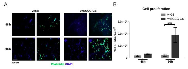
Figure 3.
Cells grown on sponges in vitro. (A) Immunohistochemical images of osteoblasts (UMR-106 cells) stained with phalloidin and DAPI. Cells were seeded and cultured on the sponges for up to 96 h. (B) Quantitative data from (A). Data are means and SDs. ** p < 0.01 (n = 3, ANOVA with Tukey-Kramer tests).

