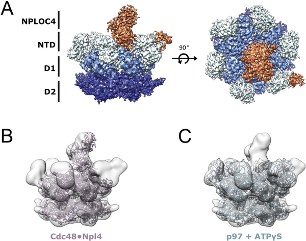
Figure 3. Cryo-EM structure of the p97-A232E·UN complex.
(A) Side (left) and top (right) views of the final sharpened density map for the p97-A232E·UN complex. Subunits and domains are colored: NPLOC4 (orange), NTDs (light blue), D1 (sky blue), D2 (navy blue). (B) Atomic model for the Cdc48·Npl4 complex (PDB:6chs, (Bodnar et al., 2018)) docked into the map for p97-A232E·UN. (C) Atomic model for ATPγS-bound wild-type p97 (PDB:5ftn, (Banerjee et al., 2016)) docked into the p97-A232E·UN complex map. See also Figure S2.

