Figure 2.
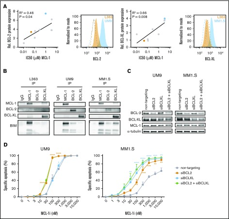
High BCL-2 or BCL-XL expression confers resistance against MCL-1i in HMCLs. (A) The correlation between BCL-2 or BCL-XL expression (fold over isotype control) as determined by flow cytometry and IC50s of MCL-1i in 9 HMCLs (RPMI-8226, NCI-H929, L363, LME-1, MM1.S, OPM-2, U266, UM1, and UM9). Representative histograms from BCL-2 or BCL-XL (filled) and isotype (dotted lines) staining of high- (blue) and low-expressing (orange) cell lines are shown, with the cell lines marked in the same colors in the adjacent correlation plot. (B) Coimmunoprecipitation of MCL-1, BCL-2, and BCL-XL and staining for BIM in L363 (MCL-1i sensitive), UM9 (MCL-1i insensitive, high BCL-2), and MM1.S (MCL-1i insensitive, high BCL-XL). (C) Protein expression 48 hours after small interfering RNA (siRNA)–mediated knockdown of BCL-2, BCL-XL, or both. (D) Specific apoptosis after 24 hours of treatment of cells from panel C with MCL-1i. Statistical significance is shown for siBCL-2 and siBCL-XL compared with the nontargeting control. *P < .05, **P < .01, ***P < .001, ****P < .0001. IgG, immunoglobulin G.

