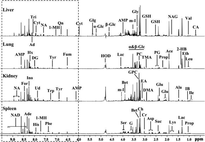
Fig. 2.
Typical 1H NMR spectra of deuterated liver, lung, kidney and spleen extracts from a low-dosed rat at 6 h p. d. The regions of δ 6.0–10.0 (in the dashed box) in the spectra were magnified 20 times in vertical expansion compared with the corresponding regions of δ 0.5–6.0. Ace acetate, Ade adenosine, Ala alanine, AMP adenosine monophosphate, Asp aspartate, Bet betaine, Ch choline, Cr creatine, Cyt cytidine, DG deoxyguanosine, DMA dimethylamine, EA ethanolamine, Eth ethanol, For formate, Fum fumarate, G glycerol, Glg glycogen, α-Glc alpha-glucose, β-Glc beta-glucose, Glu glutamate, Gly glycine, GPC glycerophosphocholine, GSH glutathione, 2-HB 2-hydroxybutyrate, His histidine, Hx hypoxanthine, m-I myo-Inositol, IB isobutyrate, Ile isoleucine, Ino inosine, Lac lactate, Leu leucine, Lys lysine, 1-MH 1-methylhistidine, NA nicotinamide, NAD NAD+, NAG N-acetylglutamate, PC phosphocholine, PG pyroglutamate, Phe phenylalanine, Prop propionate, Qn quinone, Ser serine, Suc succinate, TMA trimethylamine, Tri trigonelline, Trp tryptophan, Tyr tyrosine, Ud uridine, val valine

