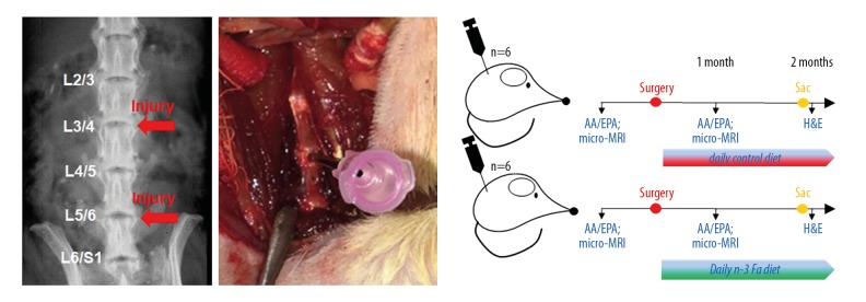
Figure 1.
Study design. Left is a radiographic image of a rat lumbar spine. The red arrows indicate the sites of disc needle injury. Middle is anterior retroperitoneal approach to the lumbar spine with exposure of 3 consecutive discs with 18-gauge needle puncture of middle IVD. Right is post-surgery: rats were assigned to either the control (oral sucrose solution) group or n-3 FA diet group. Diet supplementation occurred daily for the duration of the experiment. AA/EPA blood and micro-MRI analyses were performed in vivo. Post-surgery, histological analysis was performed. Sac – day of sacrifice; IVD – intervertebral disc; n-3 FA – omega-3 fatty acids; AA/EPA – arachidonic acid/eicosapentaenoic acid; MRI – magnetic resonance imaging.

