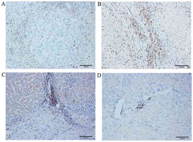
Figure 4.
Location of PD-1+ T lymphocytes in different hepatocellular carcinoma tissue regions as detected by immunohistochemical staining. Representative microscope images of the (A) nodular margin of the tumor, (B) liver tissue surrounding the tumor, (C) tumor-normal adjacent tissue junction and (D) tumor interior (magnification, ×100). PD-1, programmed cell death-1.

