Figure 3.
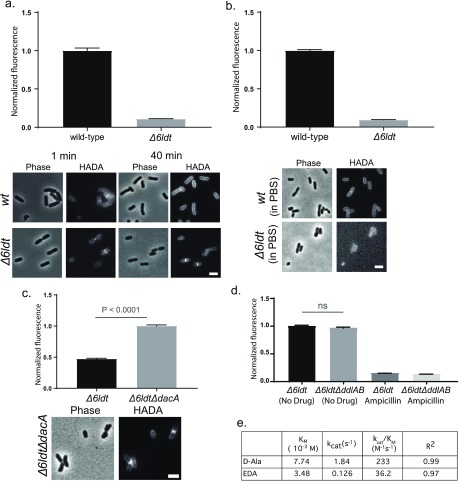
E. coli cells incorporate (F)DAAs through reactions mediated by L,D- and D,D-TPases, and not cytoplasmically. (a) HADA incorporation in E. coli BW25113Δ6LDT cells (Δ6LDT) was ∼10-fold lower than that observed in E. coli wild-type cells. Unlike wild-type cells, which reveal extensive accumulation of FDAA signal around the entire cell, FDAA labeling in the Δ6LDT strain was predominantly limited to the sites of new growth. (b) Live E. coli wild-type cells, but not Δ6LDT cells, incorporated FDAAs in a growth-independent manner. (c) Live E. coli Δ6LDTΔdacA cells accumulated significantly more HADA signal compared to Δ6LDT. (d) Live Δ6LDT and E. coli Δ6LDTΔddlAB cells, grown in M9 minimal media supplemented with DA–DA, incorporated HADA comparably; HADA incorporation was inhibited by ampicillin pretreatment. (e) Compared to D-Ala, EDA was a poor substrate for DdlBE. coliin vitro. Micrographs are adjusted for qualitative comparison only. Values in column bar graphs represent mean relative signal quantified from at least N > 100 cells. Scale bars, 2 μm.

