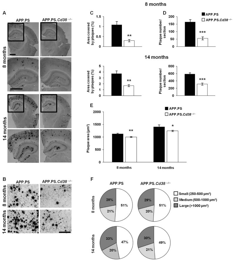
Figure 1. Loss of CD38 reduces Aβ plaque load in APP.PS mice.
Brain sections of APP.PS and APP.PS.Cd38−/− mice, 8 and 14 months of age were stained with 4G8 anti-Aβ mAb. (A) Representative images of APP.PS and APP.PS.Cd38−/− mice at 8 and 14 months of age. Lower panels represent magnifications of the selected areas in the upper panels marked by rectangles. Scale bar = 1 mm and 200 μm for higher and lower panels, respectively. (B) Representative high magnification (20×) images of 8 and 14 month old APP.PS and APP.PS.Cd38−/− mice. Scale bar = 200 μm. (C, D) Quantification of the stained area representing Aβ plaques (C) or the number of Aβ plaques (D) (**p < 0.005, ***p < 0.0005, Student’s t test). (E) Analysis of the average plaque area (*p < 0.05, **p < 0.005, Student’s t test). (F) Plaques were categorized according to their size to three groups: small (250–500 μm2), medium (500–1000 μm2) and large (> 1000 μm2) plaques. Similar distribution of the plaques in APP.PS and APP.PS.Cd38−/− mice was observed, both at 8 and 14 months of age. The quantified values shown are presented as mean ± SEM (bars) (n = 8 and 6 for APP.PS and APP.PS.Cd38−/− aged 8 months, respectively and n = 8 for APP.PS and APP.PS.Cd38−/− aged 14 months).

