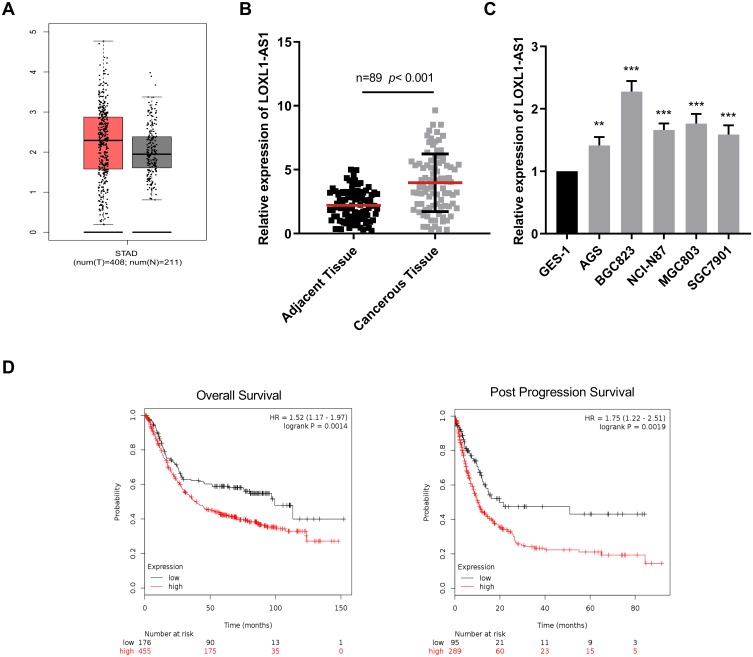
Figure 1.
The upregulation of LOX L1AS1 in GC cells was associated with the poor prognosis of patients. (A) TCGA data showed the expression level of LOXL1-AS1 was up-regulated in GC tissues. (B) qRT-PCR was used to determine LOXL1-AS1 expressions in GC tissues and adjacent normal tissues. (C) qRT-PCR was performed to measure LOXL1-AS1 expressions in normal gastric epithelial cells and 5 kinds of GC cell lines. (D) The overall survival and post-progression survival of GC patients with highly expressed and lowly expressed LOXL1-AS1 were evaluated by Kaplan–Meier method. **p<0.01, ***p<0.001.

