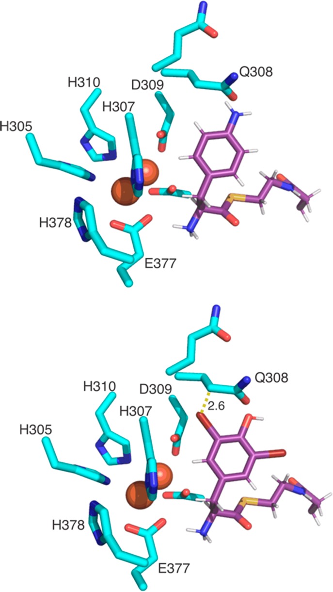
Figure 5.

CmlA active site model showing the diiron center and coordinating residues with a model of the pantetheine arm bearing either the native CmlA substrate 4-amino-Phe (upper) or 3,5-di-Br-Tyr that is not accepted as a substrate. The side chains of CmlA residues are shown in cyan sticks. The Fe atoms are shown as red spheres, and the modeled 4-amino-Phe-Pant/3,5-di-Cl-Tyr-Pant residues are shown in purple sticks.
