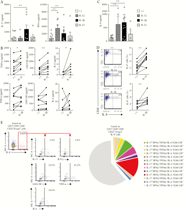
Figure 5.
Mucosal CD4+ T cells produce IL-8 in UC patients. UC mucosal CD4+ T cells were cultured with recombinant IL-12 [n = 9], IL-1β [n = 9] or IL-23 [n = 4 to 6] for 6 days. [a] IL-17 and IFN-γ, [b] TNF-α, GM-CSF, IL-6 and[ c] IL-8 secretion were measured in the culture supernatant. [d] Percentage of IL-8+CD4+ T cells after culture of UC mucosal CD4+ T cells with recombinant IL-12 [n = 10] and IL-1β [n = 8] for 6 days. [e] Ex vivo stimulation of LPMCs with PMA-ionomycin in the presence of brefeldin A for 4 h [n = 6]. Left panel: percentage of IL-17, IFN-γ, GM-CSF, TNF-α, IL-6 positive cells among CD4+CD25−Foxp3−IL-8+ T cells; right panel: pie chart depicting the co-expression of IL-17, IFN-γ, GM-CSF, TNF-α and IL-6 in CD4+CD25−Foxp3−IL-8+ T cells. [a–d] Wilcoxon signed rank test; for a and c, p < 0.01 threshold for significance to account for test multiplicity.

