
Figure 4. The dimer interface and associated lipids.
(A and B) Views of the dimer interface along (A) and perpendicular (B) to the membrane plane. The dimerisation motifs (interacting subunits coloured) are stacked along the C2-symmetry axis and formed by two copies of subunit d (red) and ATPEG1 (blue), which interacts with its symmetry-related copy, as well as ATPEG2 (green) and subunit f (yellow). Asterisks in (B) indicate positions of lipid-binding sites. (C to G) Close-ups of the lipid-binding sites indicated in (B). Bound lipids at the dimer interface identified as cardiolipin (CDL; C to E) or phospholipids modelled as phosphatidic acid (PL; F and G). Interacting residues (subunits coloured) include at least one arginine residue. Density shown as grey mesh.
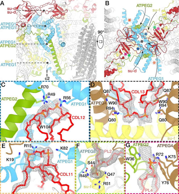
Figure 4—figure supplement 1. Architecture and dimer interface comparison between the yeast and E. gracilis ATP synthase.
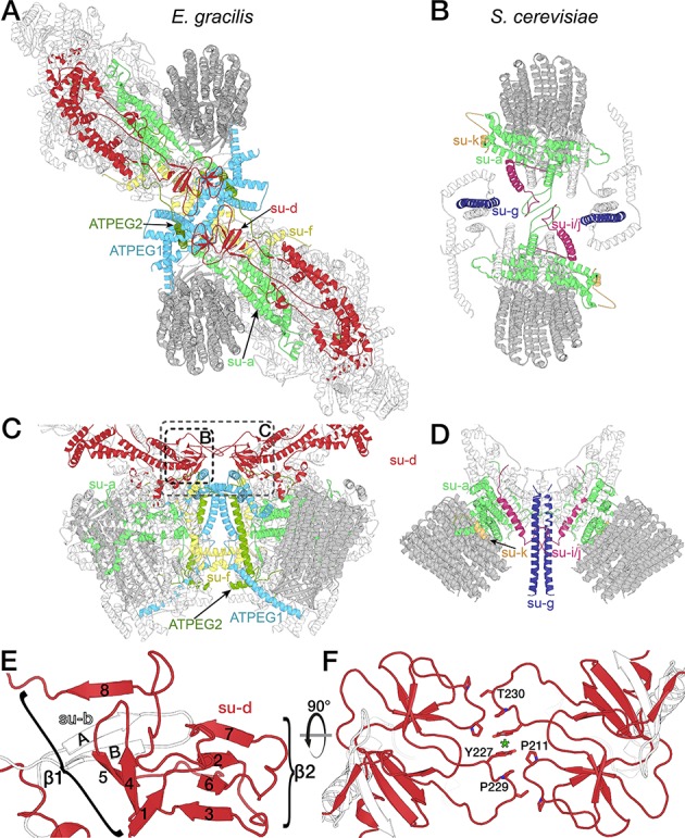
The E. gracilis (A,C) and yeast (B,D) Fo subcomplexes display different architectures. (A,C) The E. gracilis complex is held together by subunit d (red), subunit f (yellow), ATPEG-1 (light blue) and ATPEG2 (dark green). The subunit a/c-ring subcomplexes (light green and dark grey respectively) are offset along the along the Fo long axis. (B,D) The yeast ATP synthase dimer (PDB ID: 6B2Z) (Guo et al., 2017) is held together by subunit a (light green), subunit i/j, as well as subunit k (orange) and subunit g (dark blue, interaction not modelled). (E-F) Subunit d extension contributes to the dimer interface and peripheral stalk. (E) Subunit d (red) and subunit b (white) together form a ten-stranded all-β-fold at the dimer interface on the matrix side that contributes to the dimer interface. The smaller β-sheet (β2) is formed by subunit d, which adopts a ferredoxin-like fold containing a β-hairpin insertion (strands 4 an 5), which is part of the larger β-sheet (β1) that contains two β-strands (labelled A and B) of subunit b. (F) Top view of the dimer interface formed by subunit d, with interacting residues shown; green asterisk marks C2-symmetry axis.
Figure 4—figure supplement 2. Species-specific subunits and extensions form the dimer interface.
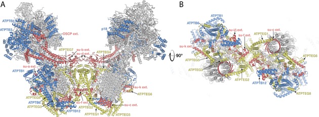
The E. gracilis ATP synthase dimer side view (A) and bottom view (B). Phylum-specific subunits previously identified in T. brucei (ATPTB1, ATPTB3, ATPTB4, ATPTB6, ATPTB12, p18) shown in blue. Species-specific subunits with no detectable homologs outside euglenoids (ATPEG1-8) are shown in yellow. Conserved subunits with homologs or close structural equivalents in yeast are shown in grey, with species-specific extensions highlighted in red.
Figure 4—figure supplement 3. Bound lipids of the dimer interface.
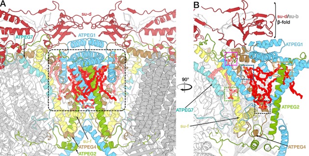
Side view (A) and cut view (B) of the dimer interface with central cavity (grey rectangle in (A)) containing bound lipids (cardiolipin red, unidentified phospholipids light pink). Subunits involved in dimerization or lipid binding are highlighted. (B) View of ATP synthase monomer cut along a plane though the C2-symmetry axis. Coloured rectangles highlight lipid binding sites at the dimer interface as shown in Figure 4C to G.