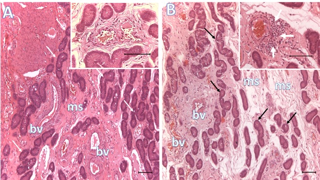
Figure 2.

Histological representative images of the endometrium. Microphotographs of endometria 6 days after BTS (control, A) or heterologous seminal plasma (SP, B) infusions prior to AI, depicting conspicuous differences in mucosal edema, vascular congestion and peri-vascular infiltration of immune cells (see inset in B, white arrows; inset in A shows lack of peri-vascular infiltration of immune cells). Arrows, uterine glands; bv, blood vessels; ms, mucosal stroma. Bar: 100 μm.
