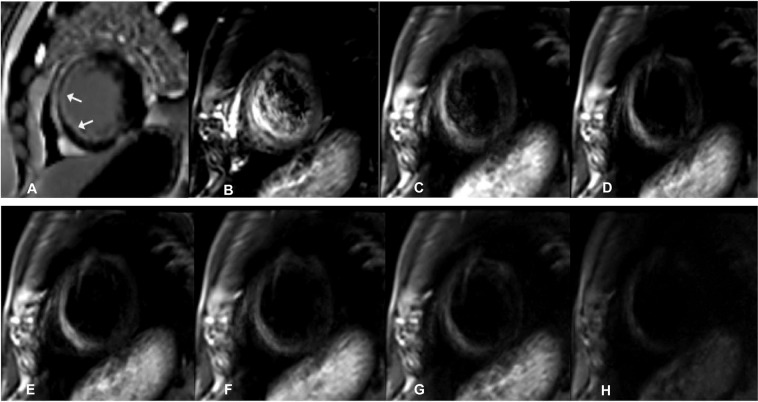
Figure 1.
A male patient aged 56 years with old myocardial infarction. (A) Magnetic resonance imaging (MRI) with delayed enhancement was performed with LGE (late gadolinium enhancement)-positive area of infarction indicated by the arrows. B-H. IVIM-DWI images of the same patient. The IVIM-DWI images were shown with b values of 0 (B), 20 (C), 60 (D), 80 (E), 120 (F), 200 (G), and 600 (G) s/mm2. Both the signal intensity of the infarcted myocardium and cardiac blood pool demonstrated high signal intensity. With the increase in the b value, the signal intensity of the blood pool decreased, with an optimal contrast at b = 120 s/mm2. IVIM, intravoxel incoherent motion; DWI, diffusion-weighted image.

