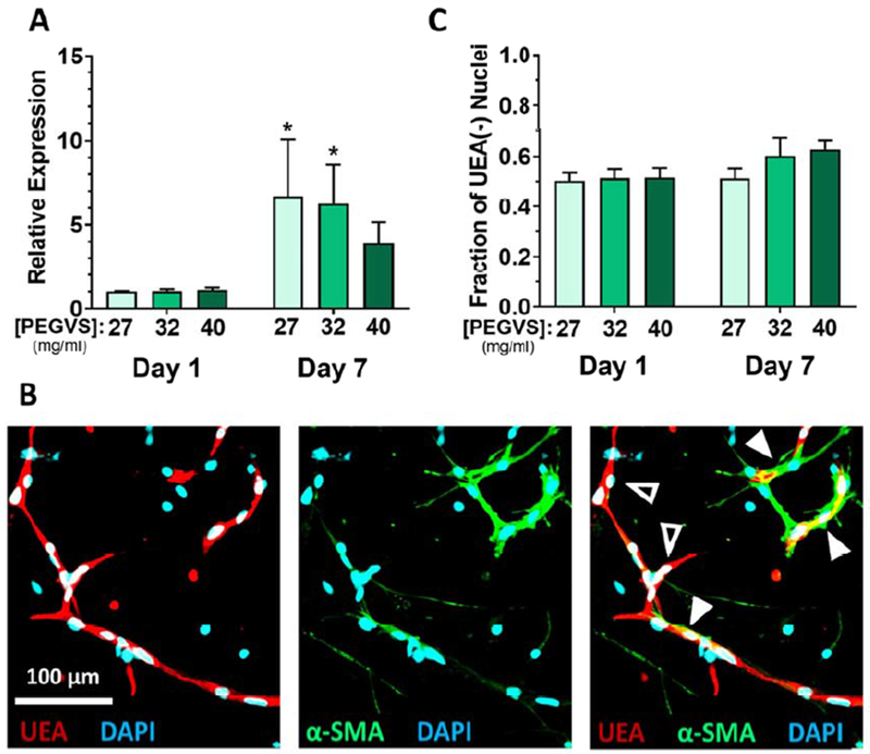
Figure 4: Smooth muscle actin is upregulated by fibroblasts in soft hydrogels.
A) Expression of α smooth muscle actin (αSMA, gene: ACTA2) was measured from RNA collected from constructs after 1 d or 7 d using real time RT-PCR. Expression levels were normalized to GAPDH in each sample and then to expression in 27 mg/mL PEGVS at 1 d (ΔΔCt method). *: p < 0.05, relative to 40 mg/mL PEGVS, N=3. B) Only fibroblasts display significant α-smooth muscle actin protein expression. Representative image in an intermediate stiffness construct showing immunofluorescent staining for (αSMA), counterstained with UEA to highlight endothelium, and DAPI. Note the tight association of αSMA+ fibroblasts with UEA+ microvessels (solid arrowheads) but lack of αSMA staining in UEA+ microvessels themselves (open arrowhead). Scale bar = 100 μm. C) The proportion of UEA(−) nuclei (representing fibroblasts) in stiff constructs after 7 d was quantified as described in the supplemental methods.

