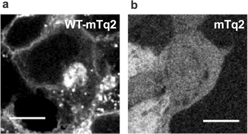
Figure 1.

Stable cell lines heterologously expressing WT-pendrin-mTq2 (a) and mTq2 (b). Fluorescence of mTq2 was imaged using a confocal microscope. The scale bars indicate 10 µm. Fluorescence images of cells expressing pendrin missense variants are provided in Supp. Fig. S3. The subcellular localizations of all pendrin constructs were qualitatively assessed and summarized in Table 1.
