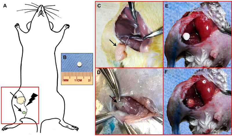
Figure 2. Schematic of the hind limb lymphadenectomy model.
(A) To induce hind limb lymphedema in mice, the inguinal fat pad (in yellow, indicated by the open arrow) and the popliteal lymph node (in green, indicated by the closed arrow) were surgically dissected. A circumferential skin incision was then made around the thigh along the dotted line, followed by hind limb irradiation. The red square indicates the field of view for images C–F. (B) Implantable pellets measured 3 mm x 3 mm. (C) Intraoperative image identifying the inguinal fat pad containing inguinal lymphatics (indicated by the open arrow), supplied by the superficial epigastric vessels. (D) Intraoperative image identifying the popliteal lymph node located within the inferior pole of the adductor thigh muscles (indicated by the closed arrow). (E) Intraoperative image of the implantable DDS placed in situ within the surgical wound. (F) Intraoperative image of the implantable DDS sutured to the fibers of the adductor thigh muscles.

