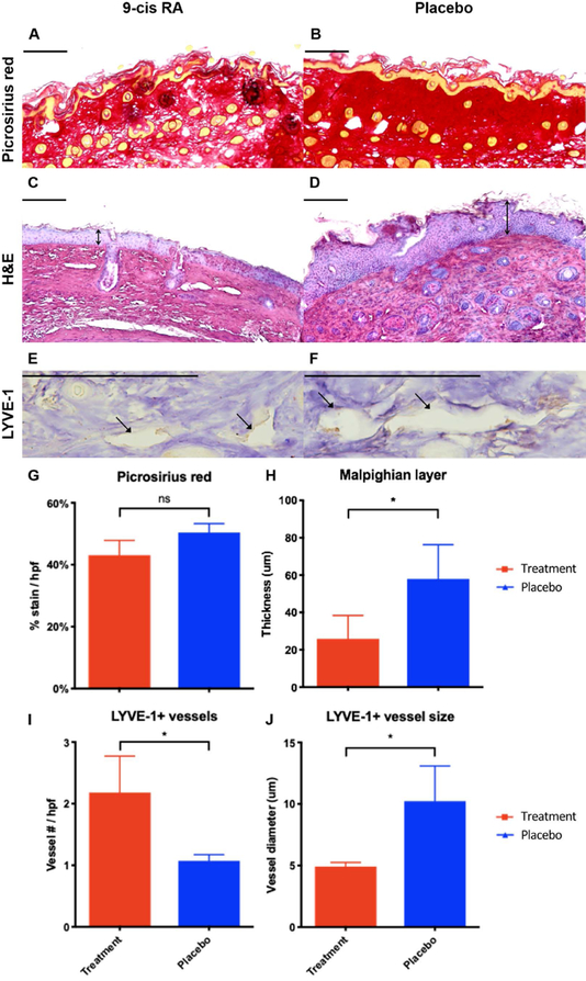
Figure 6. Sustained release 9-cis RA decreases epidermal hyperplasia, collagen deposition, and lymphatic vessel size.
(A) Representative picrosirius red stained cross-section of operated paw skin from a 9-cis RA-treated mouse. Horizontal scale bars represent 100 μm. (B) Representative picrosirius red stained cross-section of operated paw skin from a placebo-treated mouse. Note the decrease in collagen deposition in the operated paw skin of mice treated with 9-cis RA pellets. (C) Representative H&E stained cross-section of operated paw skin from a 9-cis RA-treated mouse. Horizontal scale bars represent 100 μm, vertical arrows represent Malpighian layer thickness. (D) Representative H&E stained cross-section of operated paw skin from a placebo-treated mouse. Note the decrease in hyperkeratosis and acanthosis in the operated paw skin of mice treated with 9-cis RA. (E) Representative LYVE-1 stained cross-section of operated paw skin from a 9-cis RA-treated mouse. Horizontal scale bars represent 100 μm, closed arrows indicate lymphatic vessels. (F) Representative LYVE-1 stained cross-section of operated paw skin from a placebo-treated mouse. Note the smaller lymphatic vessel diameter in the operated paw skin of mice treated with 9-cis RA. (G) Animals receiving 9-cis RA pellets showed a 15% decrease in collagen density per total tissue area compared to animals receiving placebo, indicating a reduced fibrotic response typical of lymphedema (P=0.10). (H) Animals receiving 9-cis RA pellets showed a 55% decrease in Malpighian layer thickness compared to animals receiving placebo (P=0.04), indicating reduced epidermal hyperplasia typical of lymphedema. (I) Animals receiving 9-cis RA pellets showed a 104% increase in number of LYVE-1+ lymphatic vessels compared to animals receiving placebo (P=0.04), indicating increased lymphatic density secondary to lymphangiogenesis. (J) Animals receiving 9-cis RA pellets showed a 52% decrease in LYVE-1+ lymphatic vessel diameter compared to animals receiving placebo (P=0.02), indicating preservation of the normal lymphatic architecture without luminal dilation.

