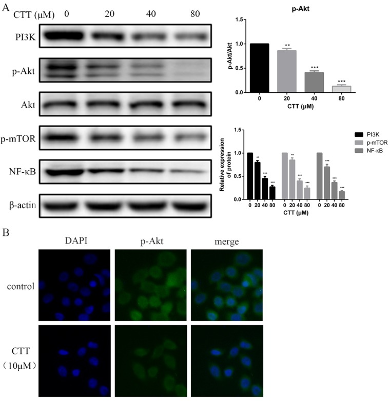
Figure 4.
CTT inhibited the PI3K/Akt and mTOR, NF-κB signaling pathways. (A) After treatment with different concentrations of CTT, images of protein expression of PI3K, p-Akt, p-mTOR and NF-κB were determined by Western blot. β-actin was used as a loading control. (B) Following treatment with or without CTT for 48 h, the expression of p-Akt in T24 cells was assessed by immunofluorescence (× 200). Each value represented the mean ± SD for triplicate samples. * P < 0.05, ** P < 0.01, *** P < 0.001, compared with control group.

