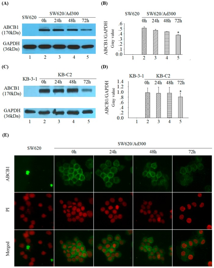
Figure 6.
(A) The expression of ABCB1 and GAPDH in SW620/Ad300 cells after treatment with 1 µM tetrandrine at 0, 24, 48, and 72 h time points. (B) representation of ABCB1/GAPDH relative ratio of A; (C) The expression of ABCB1 and GAPDH in KB-C2 cells after treatment with 1 µM tetrandrine at 0, 24, 48, and 72 h time points. (D) representation of ABCB1/GAPDH relative ratio of C; (E) Immunofluorescent staining on the influence of tetrandrine on the subcellular localization of ABCB1 in SW620/Ad300 cells at 0, 24, 48, and 72 h time points. The nuclei were stained by PI (Propidium Iodide) and shown in red.

