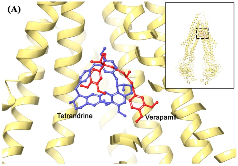
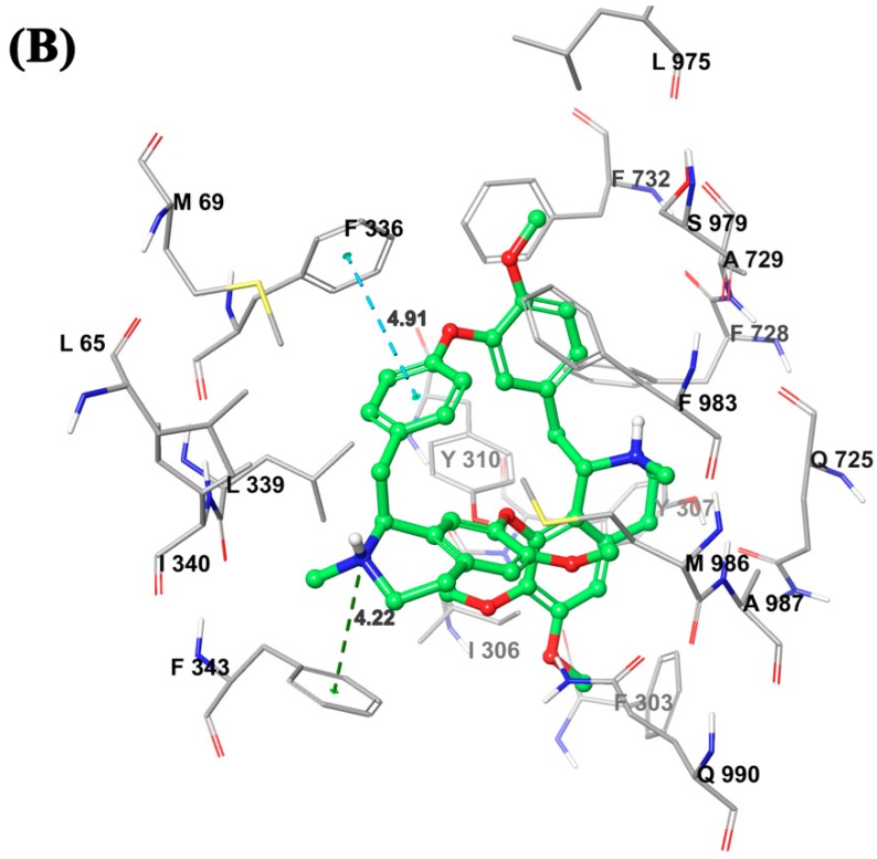
Figure 8.
(A) Comparison between binding positions of tetrandrine (blue) and verapamil (red) as ball-and-stick models. The human homology ABCB1 is depicted as yellow ribbons. (B) Induced-fit docking (IFD) predicted docked conformation of tetrandrine as ball and stick model is shown within the drug-binding site of ABCB1, with the atoms colored as carbon–green, hydrogen–white, oxygen–red, nitrogen–blue. Important amino acids are depicted as sticks with the same color scheme as above except that carbon atoms are represented in grey. Only polar hydrogens were shown. Dotted blue line indicates π-π stacking interaction, while dotted dark green line indicates cation-π interaction. Values of the relevant distances are given in Å.


