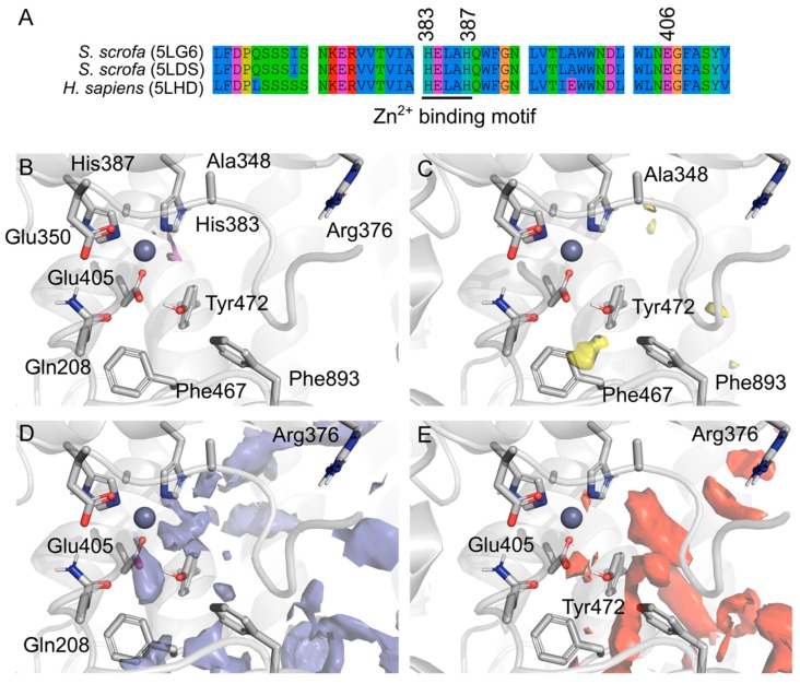
Figure 3.
(A) Amino acid sequence alignment of Porcine AMP (PDB entries 5LG6 and 5LDS) and the human homologue (5LHD), overall human and porcine AMP share 79% of sequence identity and 87% similarity, however the binding domain is conserved with punctual changes. Amino acids are colored by property and the conserved Zn2+ binding motif is underlined. SiteMap prediction of the druggable binding site near the region of the amastatin binding site from the literature. Surfaces represent the different regions in the binding pocket and are colored by property: The metal coordination (purple, (B)), hydrophobic (yellow, (C)), hydrogen bond donors (blue, (D)) and hydrogen bond acceptors (red, (E)).

