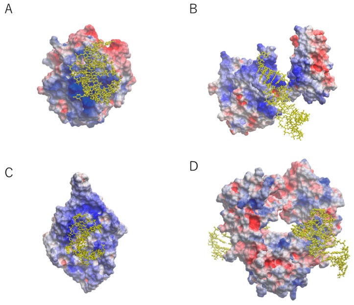
Figure 1.
Overall structure of known aptamer–protein complexes with electrostatic surface potential [5]. The RNA aptamer is represented by a yellow ball-and-stick model. (A) Aptamer–thrombin complex at 1.8 Å resolution [13]. (B) Aptamer–nuclear factor-κB complex at 2.45 Å resolution [11]. (C) Aptamer–MS2 coat protein complex at 2.8 Å resolution [12]. (D) Aptamer–Fc region of human IgG1 (hFc1) complex at 2.15 Å resolution [16]. Blue areas: positively charged; red areas: negatively charged.

