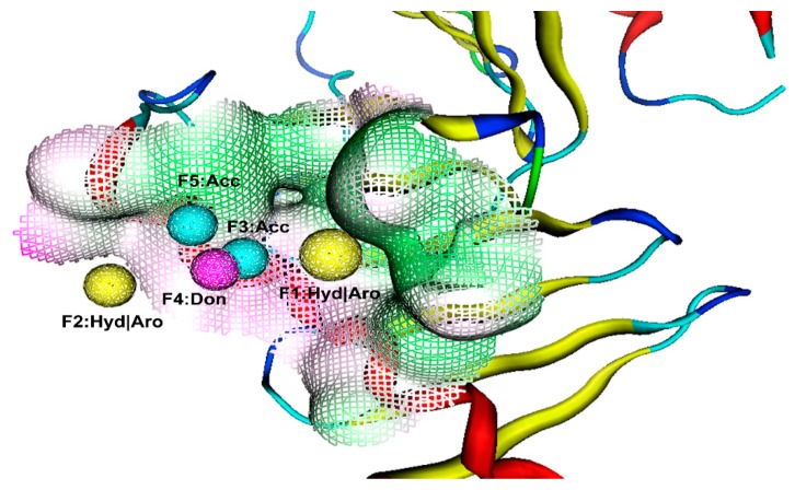
Figure 1.
The generated pharmacophore model in the binding site of PLK1-PBD. Pharmacophore features are color-coded: Yellow, two hydrophobic and aromatic features (F1 and F2: Hyd|Aro); cyan, two hydrogen bond acceptor features (F3 and F5: Acc); purple, one hydrogen bond donor feature (F4: Don). The protein backbone is shown in tube form; a reticulate pocket represents the shape of the binding site in PLK1-PBD.

