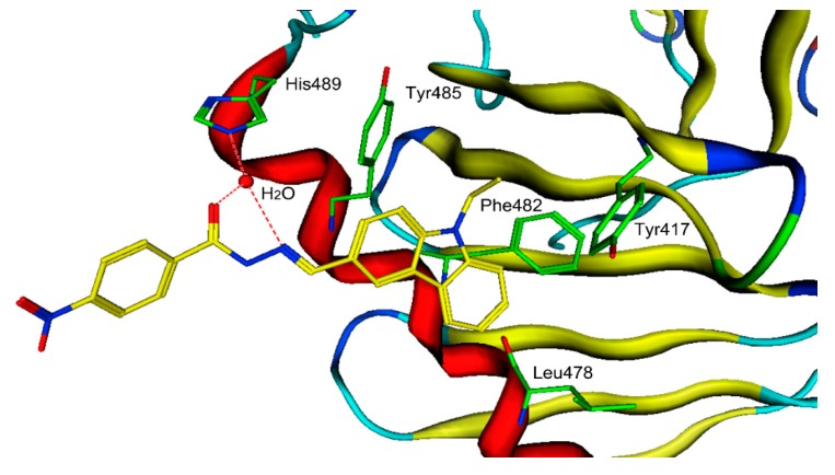
Figure 5.
The three-dimensional (3D) ligand–protein interaction diagram for the binding site of PLK1-PBD (PDB ID: 5NN2) with hit-5. The active site residues are shown in green stick form. Hit-5 is color-coded by yellow. The hydrogen-bond network with protein residues is represented by red dotted lines. The protein backbone is shown in tube form.

