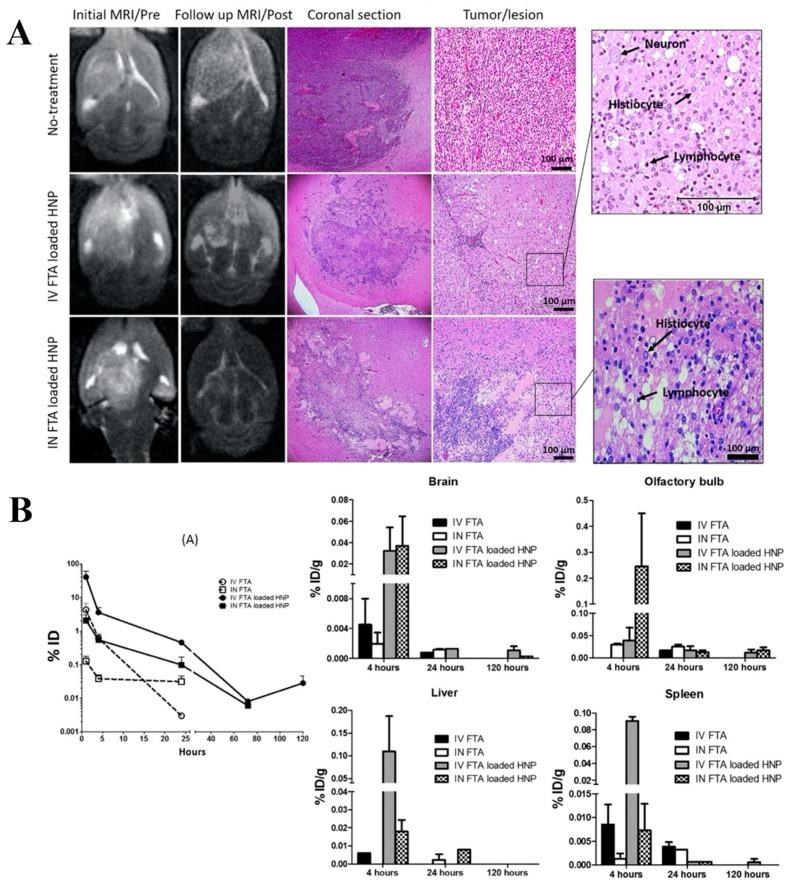
Figure 3.
Initial/pre-treatment and follow up/post-treatment MRI images of rat brains from non-treated animals or after treatments with IV or IN farnesylthiosalicylic acid (FTA)-loaded hybrid nanoparticles (HNP) formulations and their corresponding coronal brain sections stained with hematoxylin and eosin (Panel A). In the coronal brain sections, the upper panels show a dense tumor area in the right striatum of non-treated rats, whereas the middle and lower panels show cellular re-organization of tumor cells after treatment with IV or IN administered FTA-loaded HNP, respectively. Presence of inflammatory response is shown by the abundant presence of histiocytes and lymphocytes. Biodistribution study of the formulations in healthy rats (Panel B). (A) Plasma FTA concentration versus time profile for the four treatment formulations. (B) Distribution of FTA in brain, olfactory bulb, liverm and spleen of healthy rats after 4, 24, and 120 h post-administration (reproduced with permission from [123]).

