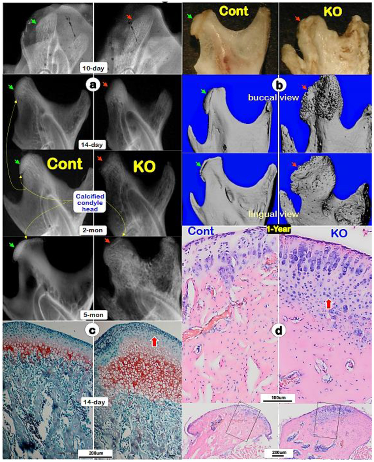
Fig. 1. Progressive changes of Dmp1 knockout (KO) condyles, which are exacerbated with age.
(a) Representative radiographs of 10-days, 14-days, 2-months and 5-months showed a largely lack of calcified condylar head with poorly formed condylar ramus in KO mice (right panels, red arrows) compared to the age matched controls (left panels; green arrows). (b) Representative 1-year-old photographs (top panels; arrows), Micro-computed tomography (μ-CT) buccal views (middle panels; arrows) and μ-CT lingual views (lower panels; arrows) displayed great expansions of KO condyle and ramus compared to the age matched controls;(c) Safranin O staining images displayed great expanded hypertrophic area and disorganized subchondral bone in a 14-day-old KO condyle (right panel) compared to a control (left panel); and(d) Toluidine blue images exhibited a continuous expansion of the KO condylar head at age of one-year (right panel) compared to the age matched control (left panel).
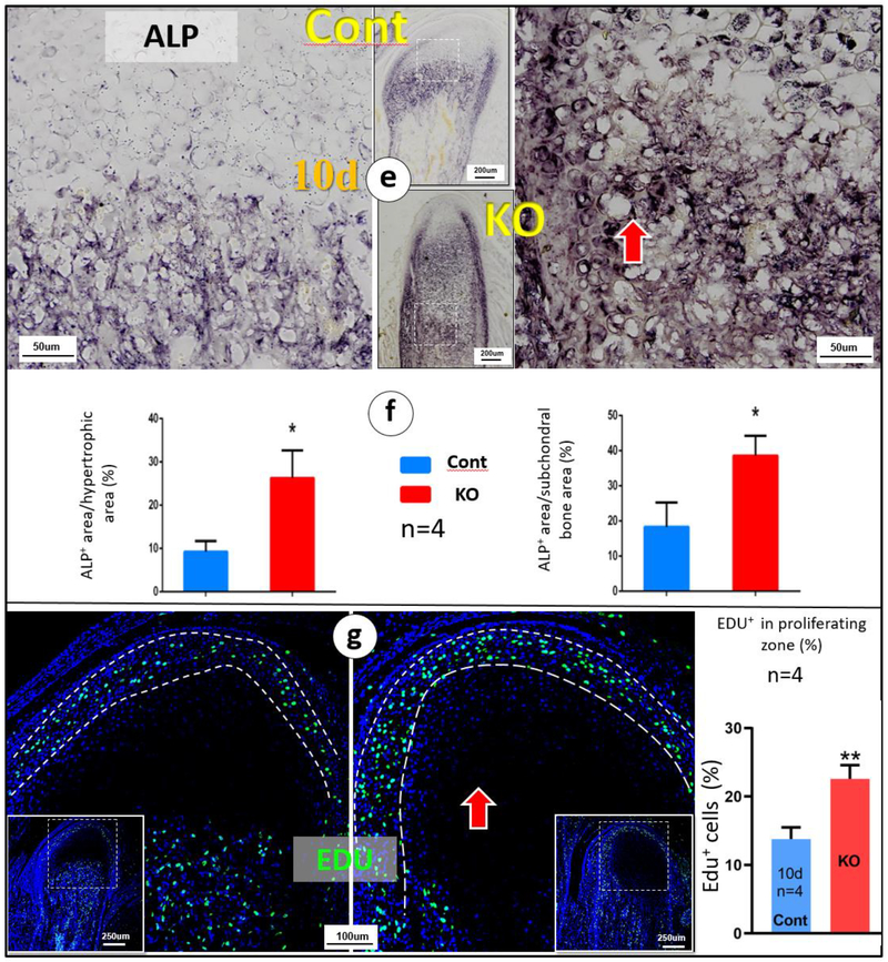
(e) Alkaline phosphatase (ALP) activity was higher in the 10-day-old KO condyle (right panel) compared to the age matched control;(f) Quantitative analyses revealed significant differences of ALP activities between the KO and control in both hypertrophic chondrocyte zone and subchondral bones (n=4; p < 0.05); (g) Representative EDU images revealed more proliferated chondrocytes in the KO condyle (right panel); a significant difference of EDU (n=4; p < 0.01).


