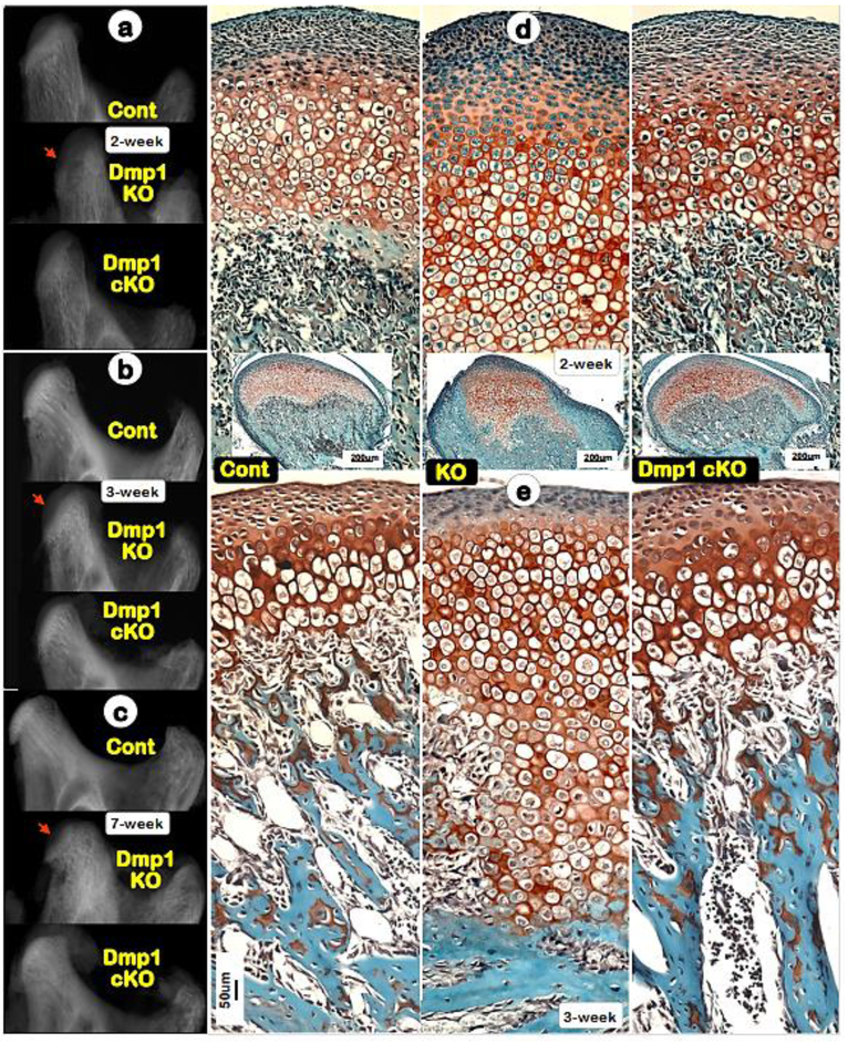
Fig. 2. Deletion of Dmp1 in hypertrophic chondrocytes results in no apparent impact on condyle morphology.
(a-c) Representative radiograph images revealed striking condyle defects in the conventional Dmp1 KO mice (middle panels), although the Col10a1-Cre; Dmp1fx/fx (Dmp1 cKO) mice display no dramatic change in its condyle (lower panels) at ages of 2-, 3- and 7-week;(d-e) Representative Safranin O stain images showed great expansion of KO cartilage layers (middle panels), whereas the cKO chondrocytes (right panels) were largely similar to those in controls of ages of 2- and 3-weeks (left panels)

