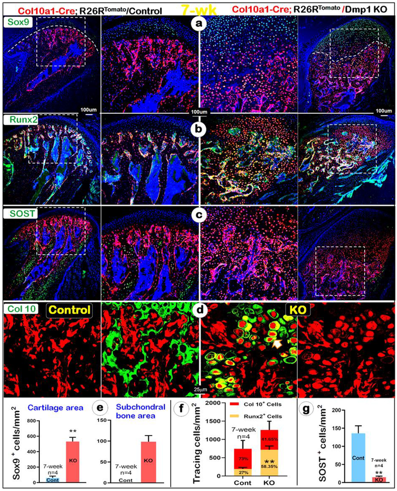
Fig. 4. An acceleration of cell trans-differentiation of hypertrophic chondrocytes to immature bone cells in the 7-week-old KO condyle.
(a-g) There was a sharp increase in the Sox9+ chondrocytes and bone cells in the KO condyle by immunohistochemistry (a, right panels); Similarly, there was a drastic increase of Col 10a1-Cre+/Runx2+ chondrocytes and bone cells in the KO condyle (b, right panels); Quantitative data analyses confirmed that these changes are statistically different in Sox9+ chondrocytes and bone cells, white line separating cartilage from bone tissue (e) and Col 10a1-Cre+/Runx2+ chondrocytes and bone cells (f); n=4, *P < 0.05, **P < 0.01. (c, g) Immunohistochemistry data showed few SOST+ cells (a marker for mature osteocyte) in the KO bone cells, indicating immature bone mass (right panel); n=4, *P < 0.05, **P < 0.01 (d) Immunohistochemistry data revealed a lack of Col 10 secretion in the KO condyle (right panel) compared to the control, in which Col 10 was secreted in the ECM (left panel).

