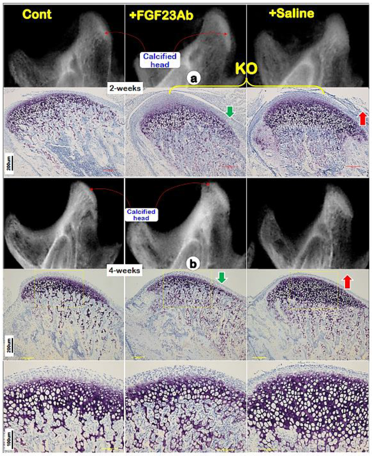
Fig. 5. Restoration of Dmp1 KO condyle defects using anti-FGF-23 antibody injections.
(a) Representative X-ray (upper middle panel) and Toluidine blue stain (lower middle panel) images of Dmp1 KO condyles showed rapid improvement of the KO condyle compared to the saline treated group (right panels) after one week treatment of anti-FGF-23 antibodies;(b) Representative X-ray (upper middle panel) and Toluidine blue stain (lower middle panels) images of Dmp1 KO condyles showed largely restorations of the KO condyle compared to the saline treated group (right panels) after three week treatment of anti-FGF-23 antibodies.

