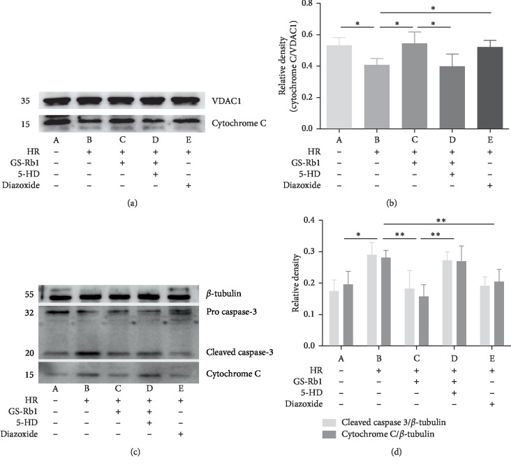
Figure 14.
Expression of apoptosis-related proteins in H9c2 cells. (a) Representative western blot illustrating the mitochondrial localization of cytochrome c. (b) Densitometric analysis of the western blot of panel (a). (c) Representative western blot illustrating the cytoplasmic localization of cytochrome c and of cleaved-caspase-3. (d) Densitometric analysis the western blot of panel (c). VDAC1 and β-tubulin were loading controls. VDAC1, voltage-dependent anion-selective channel 1; HR, hypoxia-reoxygenation; GS-Rb1, ginsenoside Rb1; 5-HD, 5-hydroxydecanoate.

