Figure 5.
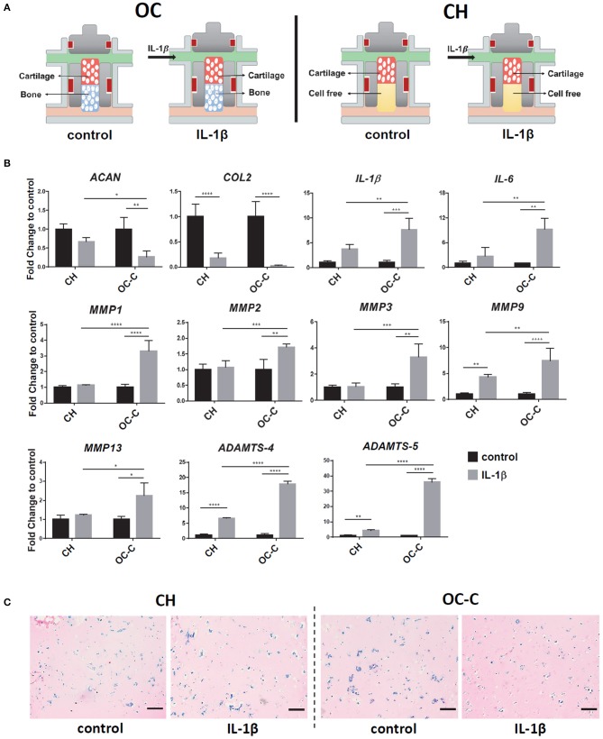
Creation of OA model in iMPCs-derived tissue chip. (A) Schematic of the process. After 28 days of differentiation and the formation of OC or CH, 1 ng/ml IL-1β was introduced into the top flow and perfused only onto the cartilage component. (B) Expression levels of anabolic factors (ACAN, COL2), proinflammatory cytokines (IL-1β, IL-6), and catabolic factors in OC or CH, with or without the treatment of IL-1β. All data are normalized to RPL13A. N = 4 per group. *p < 0.05; **p < 0.01; ***p< 0.001; ****p< 0.0001. (C) Alcian Blue staining of cartilage from OC or CH samples, with or without the treatment of IL-1β. Bar = 200 μm.

