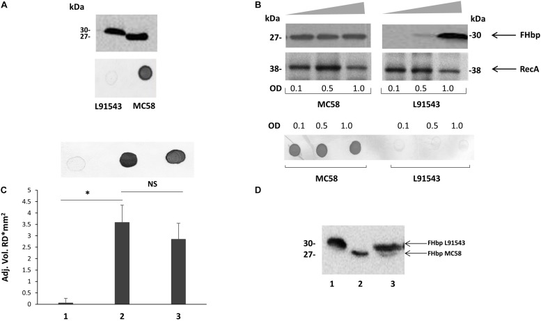
FIGURE 2.
Cleavage and surface localization of FHbp in L91543 upon transformation with MC58 fHbp. WCL (A,B,D) and whole cell suspensions from fresh plate cultures (A,C), or from broth cultures (B), analyzed by Western immunoblot and immuno-dot blot respectively with JAR4. (A) Western immunoblot (upper panel) and immuno-dot blot (lower panel) of strains L91543 and MC58. (B) Different growth phases of broth cultures of MC58 and L91543 analyzed by Western immunoblot (upper panel), including anti-recA antibody to verify loading control, and immuno-dot blot (lower panel), representative of three independent experiments. (C) Immuno-dot blot and (D) Western immunoblot: Lanes; 1, L91543; 2, MC58; 3, L91543fHbpMC58. Data are representative of five independent experiments. For (C), the reflective density was measured by a GS-800TM calibrated densitometer. The one way ANOVA followed by Dunnett’s test was employed for statistical analysis in GraphPad (V. 6). All columns represent mean ± SEM, ∗p < 0.05 vs. strain MC58.

