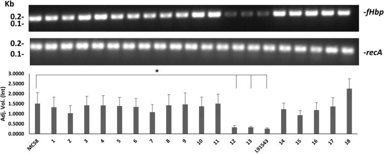
FIGURE 7.
Comparison of fHbp transcript level between strains. RT-PCR of fHbp and recA. One representative experiment of 3 is shown. Band intensity was measured using Image lab v4.0.1 and the data acquired using Linear non-threshold model (lnt). The one-way ANOVA followed by Dunnett’s test was employed for statistical analysis in GraphPad (V. 6). All columns represent mean ± SEM, ∗p < 0.05 vs. strain MC58.

