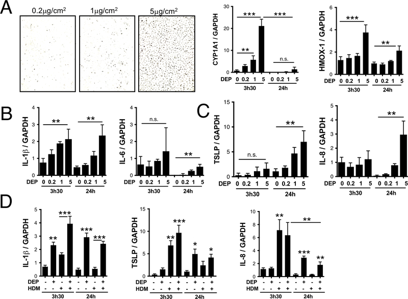
Figure 1: Delayed TSLP induction by DEP stimulated bronchial epithelial cells.
HBECs were grown in 6 well plates to 90% confluence and starved over-night before stimulation with 0.2, 1 or 5μg/cm2 of DEP (A). Media was removed and replaced with TRIZOL 3h30 and 24h later. (A) CYP1A1 and HMOX-1 mRNA levels; (B) IL-1β and IL-6 mRNA levels and (C) TSLP and IL-8 mRNA levels were determined by real time quantitative PCR and normalized to GAPDH. (D) IL-1β, IL-8 and TSLP mRNA levels following exposure to 5μg/cm2 of DEP, 25μg/ml of HDM or both (data compiled from two separate experiments).

