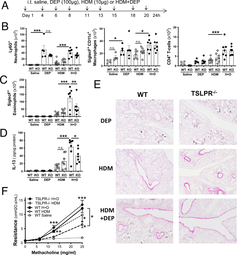
Figure 2: TLSP contributes to HDM+DEP induced eosinophilia but is not required for HDM+DEP induced AHR.
(A) Experimental protocol. (B) BALF levels of Gr-1+ neutrophils, SiglecF+CD11c+ macrophages, CD4+ T-cells, and SiglecF+ eosinophils (C) were assessed by flow cytometry (n=4–7 mice/group; 1 way-ANOVA, * p<0.05 ** p<0.01, *** p<0.001, n.s.= not significant). (D) IL13 levels were assessed in lung homogenates by ELISA and normalized to total lung protein levels. (E) Representative PAS stained lung sections. (F) Airway resistance was measured a day after the last challenge using FlexiVent (n=7–13 mice/group from two separate experiments; 2-way ANOVA, ** p<0.01, *** p<0.001).

