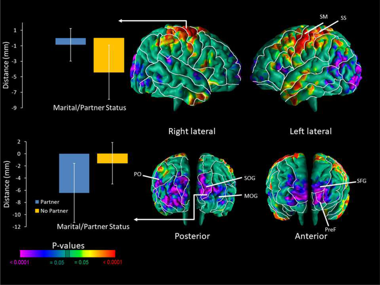
Figure 2. Correlations of Cerebral Surface Measures with Partner Status.
Maps are shown for partner status correlations with morphological measures of the cerebral surface. The p-values are adjusted for multiple comparisons with FDR. We found significant inverse correlations of local brain volumes in the neonates based on maternal partner status in the prefrontal region and diffuse across the occipital lobe of both hemispheres, and the angular gyrus of the left hemisphere. There were positive correlations of local volume in the fronto-parietal and inferior temporal regions of both hemispheres. A sample of the average local volumes (or, more accurately, distances in mm from the most significant corresponding point on the surface of the template brain in the region denoted) are displayed in bar charts for the SM and SOG of the right hemisphere. Abbreviations: PreF – Prefrontal; MOG – Middle Occipital Gyrus; PO – Parieto-occipital; SM – Somatomotor; SS – Somatosensory; SFG – Superior Frontal Gyrus; SOG – Superior Occipital Gyrus.

