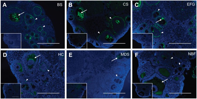
Figure 4.
Ethanol-Formalin-Glacial Acetic Acid, Carnoy’s Solution, and Bouin’s Solution produce optimal HABP signal at low magnification in the ovary. Representative images of HABP-stained ovarian tissue fixed in (A) Bouin’s Solution (BS), (B) Carnoy’s Solution (CS), (C) Ethanol-Formalin-Glacial Acetic Acid (EFG), (D) Histochoice (HC), (E) Modified Davidson’s Solution (MDS), and (F) 10% Neutral Buffered Formalin (NBF). For all images, DNA is shown in blue and HA is shown in green. HA shows greatest localization in the follicular fluid of antral follicles (arrow) as well as in the layer of theca cells immediately surrounding growing follicles (arrowhead). Fixation with EFG, Carnoy’s Solution, and Bouin’s Solution produced greater HA signal brightness. Negative controls showing sections treated with bovine testicular hyaluronidase, which degrades HA and abrogates HABP signal, are shown in the insets. Scale bars are 400 μm.

