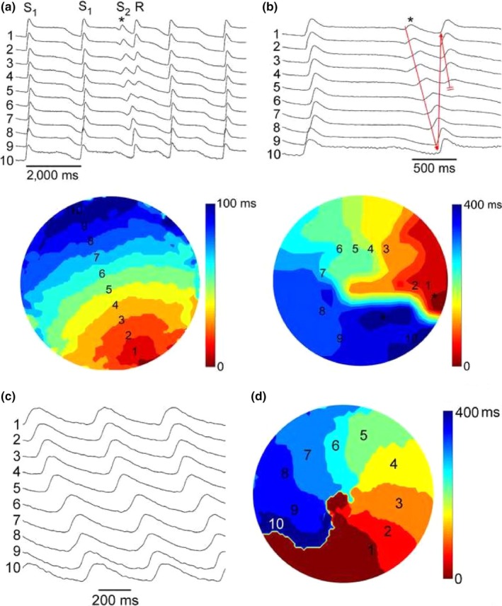
Figure 2.

Panels (a) and (b): Initiation of a single spiral wave reentry by an S1S2 protocol. The figure illustrates the last two beats of a series of 10 S1S1 stimulations at 0.5 Hz from a site close to the bottom of the optical field. The preparation was perfused with 4 nM AP‐A, resulting in prolongation of FCaiD85 to 1,500–1,600 ms. An S2 stimulus was applied to a site close to the right edge of the optical field at a CL of 1,250 ms (marked by asterisk) and induced a single reentrant spiral wave (R). Panel (a) shows 10 selected FCai potentials to illustrate the pathway of the FCai propagation wave of the S2 stimulus. Panel (b) illustrates an expanded view of the last S1 stimulus, the S2 stimulus, and the single spiral wave reentry. The isochronal map of both the S1 and S2 stimuli is shown underneath. The S1 map is drawn at 10 ms isochrones while the S2 map is drawn at 40 ms isochrones. The S1 stimulus resulted in a smooth propagation wave from the bottom to the top of the optical field at an average conduction velocity of 15 cm/s. On the other hand, the S2 stimulus failed to propagate to the bottom right half of the optical field resulting in a line of functional block that extended from the right edge to the middle of the optical field. The FCai wave front circulated around the left side edge of the line of block. However, the circulating wave front failed to propagate through the bottom side of the line of block, thus resulting in a single reentrant cycle. The expanded recordings in panel (b) illustrate both propagation of the circulating wave front (red arrow) and its final failure of propagation at the lower side of the line of functional block (double red bars). Panels (c) and (d) illustrate FCai signals and an isochronal map of sustained spiral wave reentry that was obtained 30 s following the recording in Panel (a) utilizing a similar S1S2 protocol. The spiral rotated at a CL of 400 ms around a 2 mm diameter core in the center of the optical field at approximately the same site as the left side end of the line of functional propagation block during the initiation of reentry in figure (a)
