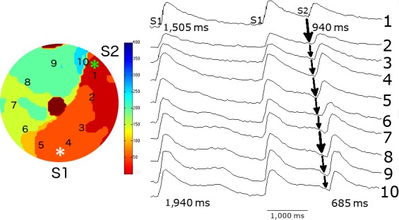
Figure 6.

Initiation of spiral wave reentry by an S1S2 stimulation protocol from the monolayer with a central anatomical obstacle. The middle panel illustrates the last two beats of a series of 10 S1S1 stimuli at 0.5 Hz and their position is shown in the right panel. An S2 stimulus (marked by asterisk) was applied to a site close to the zone of functional spatial dispersion of FCaiD85 and induced a single reentrant spiral wave. The selected FCai signals of S2 stimulus illustrate the pathway of the FCai propagation wave. The isochronal map of the S2 stimulus is shown on the left panel. The S2 stimulus failed to propagate in a counterclockwise direction because of a continuous line of block that includes the central anatomical obstacle and a functional zone of block. The wave front instead circulated in a clockwise direction around the central anatomical obstacle but failed to propagate through the functional line of block at the 1 o'clock zone, thus resulting in a single reentrant cycle. The figure illustrates the possible electrophysiological mechanism of the failure of the counterclockwise wave front to break through the line of functional block. The FCaiD85 of the two S1 signals on each side of the line of functional block were 1,505 and 1,940 ms. The duration of the closely coupled S2 signal at both sites showed significant shortening as expected. However, there was relatively more shortening of the FCaiD85 bottom signal. This resulted in a shorter duration of the FCaiD85 bottom S2 signal on the left side of the functional block compared to the FCaiD85 top S2 signal on the right side of the line of block. Repetitive reentry would have required a longer duration of the FCaiD85 S2 signal of the left side of the line of functional block compared to the signal on the right side of the block
