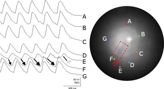
Figure 8.

The mechanism of spontaneous termination of the spiral wave reentry. The figure illustrates seven FCai signals from selected pixels around the spiral wave front (marked by different colors on the fluoroscopic image of the monolayer and labeled A to G). The recording illustrates the sequential rotation of the FCai wave front around the anatomical obstacle. The termination of the spiral wave reentry occurred at a site located between the E and F traces (marked by a line with double cross bars). The gradual lengthening of the arrows drawn between the E and F traces represents gradual slowing of propagation of the FCai wave front between these two sites before propagation failure terminated the reentrant activation. The conduction delay and terminal block of reentrant activation took place in the normal section of the monolayer outside the fixed anatomical obstacle. Thus, termination of the circulating wave front has to occur at a continuous line of block that consists of the anatomical fixed obstacle and a functional line of block that extends the electrophysiological barrier to the edge of the monolayer
