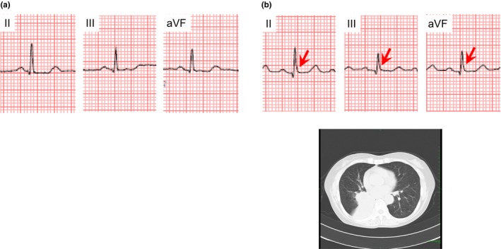
Figure 4.

A 61‐year‐old woman who presented J waves coincidently with development of lung cancer. (a) No J wave was detected before the hospital admission. (b) J waves with slurring morphology (arrows) appeared in leads II, III, and aVF, when lung cancer contacted the heart on the hospital admission
