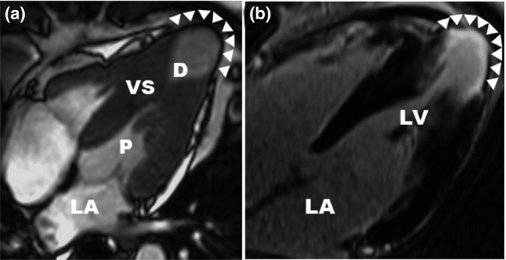
Figure 1.

cardiac magnetic resonance (cited from Rowin et al) (a) CMR demonstrates medium‐size apical aneurysm (3.2 cm; arrowheads) with associated hourglass shaped LV chamber producing distinct proximal (P) and distal (D) chambers. (b) LGE localized to the aneurysm rim (arrowheads)
