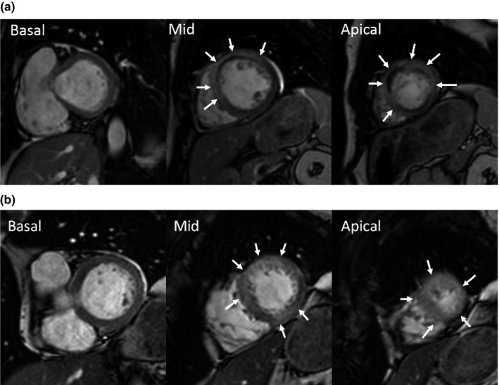
Figure 1.

Contrast‐enhanced cine images from cardiac magnetic resonance in three short axis slices where the myocardium at risk (MaR) is indicated by arrows. In patient (a) with ST‐elevation in aVL there is MaR in the apical lateral segment but no inferior MaR. Conversely, in patient (b) without STE in the aVL there is MaR in the midventricular and apical inferior segment, corresponding to a large wrap‐around of the LAD coronary artery. However, there is no lateral MaR in the midventricular segment
