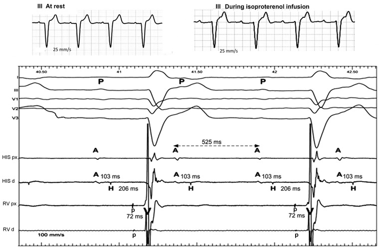
Figure 2.

His bundle recordings during Isoproterenol infusion precipitating 2:1 AV block at a sinus rate of about 115/min (PP = 525 ms). Top: Lead III ECG (recorded with an electrocardiograph at 25 mm/s) before and after isoproterenol infusion. Under the slow ECG on top, the ECG leads I, III, and V1–V3 were recorded at 100 mm/s. A = atrial deflection, H = His bundle potential, V = ventricular potential, RV = right ventricular, p = right bundle branch or Purkinje potential, P = P wave, pV = interval from p to V, His px = channel recording from proximal His bundle, His d = channel recording from distal His bundle, RVp = channel recording from proximal RV, RVd = channel recording from distal RV. This represents classic infra‐Hisian AV block. During AV block, the p potential is absent indicating block between the His bundle and the p potential. See text for details.
