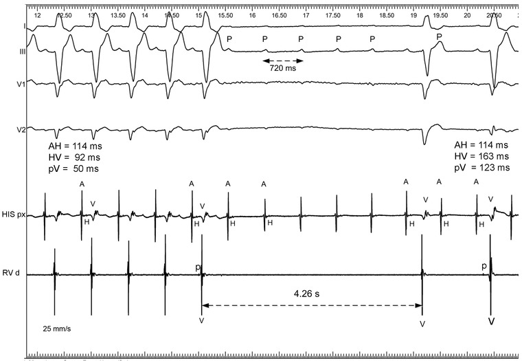
Figure 3.

Paroxysmal AV block induced by isoproterenol infusion at a sinus rate of about 83/min (PP = 720 ms). ECG leads I, III, V1–V3. A = atrial deflection, H = His bundle potential, V = ventricular potential, RV = right ventricular, p = Purkinje or right bundle potential, P = P wave, pV = interval from p to V, His p = channel recording from proximal His bundle, RVp = channel recording from proximal RV. This form of AV block is different from that in Figure 2 because the Purkinje p potentials were recorded during AV block so that the block appeared distal to the p or right bundle branch potential. All the AH intervals are constant and indicate supraventricular conduction. The last two ventricular beats are probably ventricular escape beats. They are most probably not conducted and are unlikely to represent ventricular fusion. See text for details
