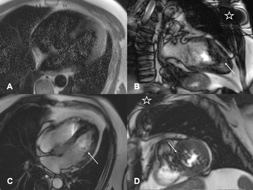
Figure 1.

Cardiac MRI images in a patient with paroxysmal atrial fibrillation (patient 1). Long‐axis four‐chamber view HASTE sequence (A) with diffuse grainy hyperintense susceptibility artifacts which were detected in every second image slice. (B and C) Long‐axis two‐ and four‐chamber view and a midventricular short‐axis view (D) SSFP cine sequence with prominent dark rim artifacts affecting the apex, apical septum, and lateral left apical and midventricular myocardium (arrow; B–D). Star indicating the left pectoral location of the ILR. HASTE = half‐Fourier acquisition single‐shot turbo‐spin‐echo; SSFP = steady‐state free precession; ILR = implantable loop recorder.
