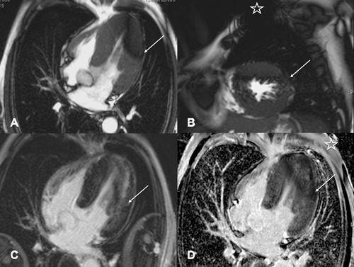
Figure 2.

Cardiac MRI images in a patient with known history of Anderson‐Fabry disease and myocardial involvement with left ventricular hypertrophy (patient 2). Long‐axis four‐chamber view (A) and short‐axis (B) view in SSFP cine sequence with dark rim artifacts (arrow) affecting the apical to mid ventricular lateral left chamber wall. LGE 10 minutes after a double dose of gadobutrol i.v. (C and D) shows discrete hyperintense rim artifacts in the left lateral wall in Turbo‐FLASH (fast low angle shot) sequences with variable time to inversion (C; arrow) above a diffuse midmyocardial area of pathological LGE which is typical for cardiac involvement with Anderson‐Fabry disease. Phase‐sensitive inversion recovery sequence (D) shows darker rim like artifacts in the same region (arrow). Star indicates ILR on left chest wall (B and D). SSFP = steady‐state free precession; LGE = late gadolinium enhancement; ILR = implantable loop recorder.
