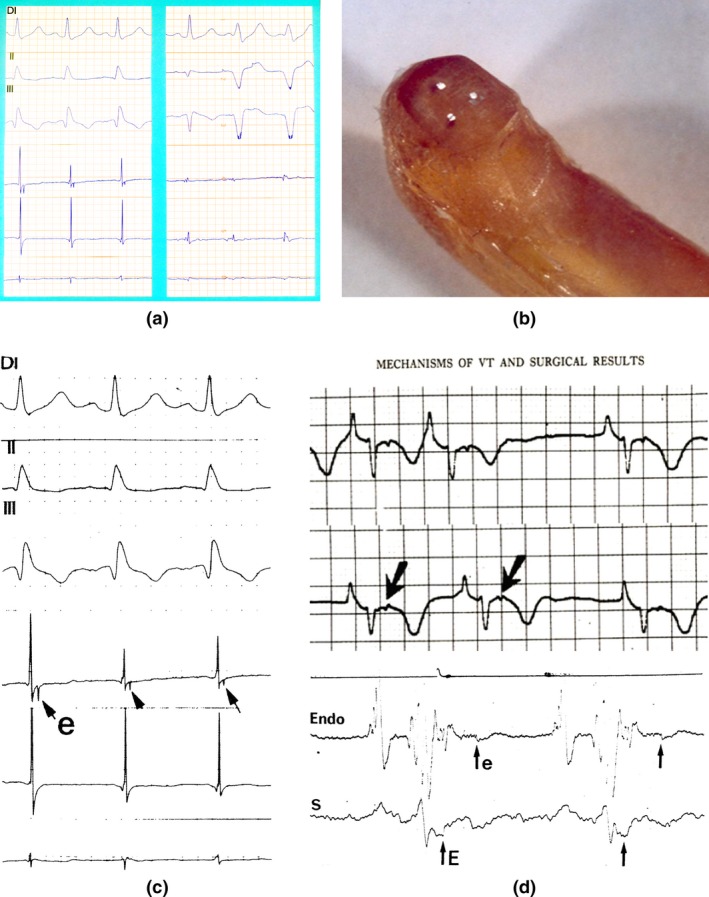
Figure 1.

(a) Left: First recording of an Epsilon wave on the border zone of a diaphragmatic scar in a patient with an old myocardial infarction (insert arrow). Right: Epicardial potentials recorded on the scar tissue (insert arrow). (b) The electrodes are separated by 1.5 mm. Handmade instrument in Epoxy resin and platinum electrodes (With permission from Dr. Guy Fontaine, 2010). (c) First presentation of an epicardial Epsilon wave (With permission from Dr. Guy Fontaine (Fontaine et al., 1977)). (d) First presentation of an Epsilon wave on Holter‐ECG and on the endocardium (With permission from Dr. Guy Fontaine (Fontaine et al., 1977))
