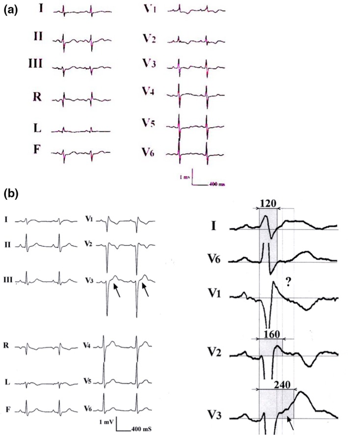
Figure 3.

(a) An incomplete RBBB. (b) An example of more than incomplete right bundle branch block recorded in a young arrhythmogenic right ventricular dysplasia human with palpitations. The interesting aspect of this tracing was that the Epsilon wave is barely visible on a standard ECG. However, using the double amplitude recording it was possible to disclose late potentials up to 240 ms after the beginning of the QRS in some leads as opposed to lead I or V6 with a duration of 120 ms. The ‘?’ sign stresses the limit of Epsilon wave recognition on a single lead (With permission from Dr. Guy Fontaine (Fontaine et al., 2017))
